GENTAUR - Research solutions for the academic, biotechnology and pharmaceutical industries
![]()
|
|
Mito Casp™
A simultaneous dual parameter Assay For: Mitochondrial Membrane Potential Detection & Caspase (poly, 3/7, 8, 9, 1) Activity Patent Pending*
|
| Key Benefits: |
- Simultaneous detection of mitochondrial membrane potential and caspase activity.
- Readout - Flow cytometry, Fluorescent plate reader, Fluorescent microscopy .
- Reliable: Yields both quantitative and qualitative results. Gives a strong positive signal.
- The kit can be used in conjunction with other antibodies or stains.
- Ease Of Use: No need to make cell lysates or run western blots.
- Cell Permeable Reagents.
|
| Background: |
Caspase Detection
Apoptosis is an evolutionarily conserved form of cell suicide, which follows a specialized cellular process. The central component of this process is a cascade of proteolytic enzymes called caspases. These enzymes participate in a series of reactions that are triggered in response to pro-apoptotic signals and result in cleavage of protein substrates, causing the disassembly of the cell (1).
Caspases have been identified in organisms ranging from C. elegans to humans. The mammalian caspases play distinct roles in apoptosis and inflammation. In apoptosis, caspases are responsible for proteolytic cleavages that lead to cell disassembly (effector caspases), and are involved in upstream regulatory events (initiator caspases). An active caspase consists of two large (~20 kD) and two small (~10 kD) sub units to form two heterodimers, which associate in a tetramer (2-4). As is common with other proteases, caspases are synthesized as precursors that undergo proteolytic maturation, either autocatalytically or in a cascade by enzymes with similar specificity (5).
Mitochondrial Membrane Potential Detection
The loss of mitochondrial membrane potential is a hallmark for apoptosis. The mitochondrial permeability transition is an important step in the induction of cellular apoptosis. During this process, the electrochemical gradient across the mitochondrial membrane collapses. The collapse is thought to occur through the formation of pores in the mitochondria by dimerized Bax or activated Bid, Bak, or Bad proteins. Activation of these pro-apoptotic proteins is accompanied by the release of cytochrome c into the cytoplasm (11-14). |
| Assay Principle: |
Caspase (poly, 3/7, 8, 9, 1) & Mitochondria Membrane Potential Detection - MitoCasp
Caspase enzymes specifically recognize a 4 amino acid sequence (on their substrate) which necessarily includes an aspartic acid residue. This residue is the target for the cleavage reaction, which occurs at the carbonyl end of the aspartic acid residue(6). Caspases can be detected via immunoprecipitation, immuno-blotting techniques using caspase specific antibodies, or by employing fluorogenic substrates which become fluorescent upon cleavage by the caspase. MitoCasp uses a novel approach to detect active caspases (7-9). The methodology is based on carboxyfluorescein (FAM) labeled fluoromethyl ketone (FMK)-peptide inhibitors of caspases. These inhibitors are cell permeable and non-cytotoxic. Once inside the cell, the inhibitor binds covalently to the active caspase (10). Cells that contain bound inhibitor can be analyzed by flow cytometry or fluorescence microscopy.
Gentaur utilizes a cationic dye to visualize mitochondrial membrane potential (15-17). The cationic dye is cell permeable and has a strong fluorescent signal in the red region and exhibits low membrane potential independent (non specific) binding and toxicity. In healthy cells the cationic dye is accumulated by the mitochondria in proportion to the DeltaPsi (membrane potential). In most cell lines, accumulation of the cationic dye in the mitochondria results in a higher fluorescence intensity. In apoptotic cells, where the mitochondrial membrane potential is compromised, the cationic dye does not accumulated in the mitochondria and these cells exhibit a lower fluorescence signal. Utilizing these two reagents in combination Caspase activity and mitochondrial membrane potential can be analyzed simultaneously. Citations Identification of single-domain, Bax-spec |
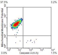
(A)
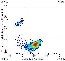
Jurkat cells were stimulated with Staurosporine for 3 hours (B) or DMSO (A). The cells were then stained with the MitoCasp kit according to the protocol. The cells were then washed twice and analyzed by flow cytometry: Ex:488nm Em: FL1 and FL2.
Fig A. Healthy cells show a strong red fluorescence indicating intact mitochondria and no green fluorescence, indicating no active caspases.
(B)
Fig B. Apoptotic cells show a loss of red fluorescence (y axis) indicating loss of mitochondrial membrane potential and positive green fluorescence (x axis) indicating active caspases.
| Kit contents |
- Mitochondrial membrane potential cationic dye
- Caspase Detection reagent (Poly Caspase , Caspase 3/7, Caspase 1, Caspase 8 or Caspase 9)
- 10X Wash Buffer
- 1X Dilution Buffer
|
|