full screen background image

GENTAUR - Research solutions for the academic, biotechnology and pharmaceutical industries


Mito Flow™ 
Mitochondria Membrane Potential detection for Flow Cytometry

Key Benefits:
  • Cell Permeable, Easy One Color Assay for Flow Cytometry.
  • Can be used with both suspension and monolayer adherent cell lines.
  • Adaptable for High Throughput format.
  • Compatible with other antibodies or stains. For example flourescent protein expression vectors.
  • Applications - Flow Cytometry.
Assay Principle

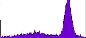
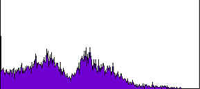
Gentaur’s Mito Flow assay utilizes a cationic dye to visualize mitochondrial membrane potential (5-7). The Mito Flow reagent is a cell permeable cationic dye that has a strong fluorescent signal and exhibits low membrane potential independent (non specific) binding and toxicity. In healthy cells the Mito Flow reagent is accumulated by the mitochondria in proportion to the DeltaPsi (membrane potential). In most cell lines, accumulation of the Mito Flow reagent in the mitochondria results in a higher fluorescence intensity. In apoptotic cells, where the mitochondrial membrane potential is compromised, the Mito Flow reagent does not get accumulated in the mitochondria and these cells exhibit a lower fluorescence signal.

MITO


(A)
Figure 1. Jurkat cells were stimulated with DMSO for 3 hours. Cells were then stained by Mito Flow and analyzed by Flow Cytometry

MITO


(B)
Figure 1. Jurkat cells were stimulated with Staurosporine for 3 hours. Cells were then stained by Mito Flow and analyzed by Flow Cytometry


Kit contents:
  • Vial of Mito Flow Dye Part#4004
  • Bottle of 10X dilution buffer Part#3004

Product Catalog # Size*
High Throughput Screen for Mitochondria Membrane Potential Kit Flo200-2 100
High Throughput Screen for Mitochondria Membrane Potential Kit Flo200-3 500