
GENTAUR - Research solutions for the academic, biotechnology and pharmaceutical industries
JC-1 Mitochondrial Membrane Potential Detection Kit ™ |
||
| Key Benefits: | ||
|
||
| Assay Principle | ||
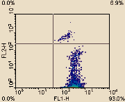
Detection of the mitochondrial permeability transition event provides an early indication of the initiation of cellular apoptosis. This process is typically defined as a collapse in the electrochemical gradient across the mitochondrial membrane, as measured by the change in the membrane potential (YD). Loss of mitochondrial (YD) is indicative of apoptosis and can be detected by a unique fluorescent cationic dye, 5,5',6,6'-tetrachloro-1,1',3,3'-tetraethyl- benzamidazolocarbocyanin iodide, commonly known as JC-1. This dye has been incorporated into the user-friendly kit for the simple and reproducible detection of the membrane potential (YD) event in apoptotic cells. The kit has been formatted for use on Flow cytometers, Fluorescent plate readers and Fluorescent Microscopes |
||

|


